Frequently Asked Questions About Canine Proptosis
While many serious conditions that affect dogs may appear so subtle as to go unnoticed for a long time, canine proptosis makes itself startlingly evident right away. This condition, in which the eye of a dog may seem to pop partially or completely out of its socket, can lead to blindness without prompt veterinary care.
You'll feel better prepared to deal with a case of canine proptosis once you know a little about what it is, why it happens, and what your veterinarian can do to fix the problem. Take a look at some answers to frequently asked questions about canine proptosis.
What Does Canine Proptosis Involve?
Canine proptosis involves a sudden forward shift in a dog's eyeball. The eyeball may appear to jut halfway out of its orbit (or socket), making the affected eye usually large and bulbous. Without proper eyelid protection and moisture retention, the cornea and/or conjunctiva of the eye may also become swollen and inflamed.
In extreme cases of proptosis, the eyeball may have popped completely out of the eye, with only its connective tissues keeping it attached to the socket. Proptosis may reduce blood supply and nerve function to the eye that results in permanent vision loss, especially without aid of immediate treatment.
Why Does Canine Proptosis Occur?
Most cases of canine proptosis occur as a result of a traumatic injury . A blow to the head or near the eye socket can cause the eyeball to pop out of its socket. A car impact or a fight with another animal can produce proptosis among other injuries.
Any condition that causes excessive pressure to build up in the eye or at the back of the eye socket can force the eyeball outward. A tumor inside the eye socket may also produce proptosis. Even too much pressure from a collar or similar neck restraint can lead to proptosis in one or both eyes.
Which Animals Have an Elevated Risk for Canine Proptosis?
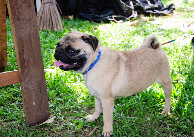
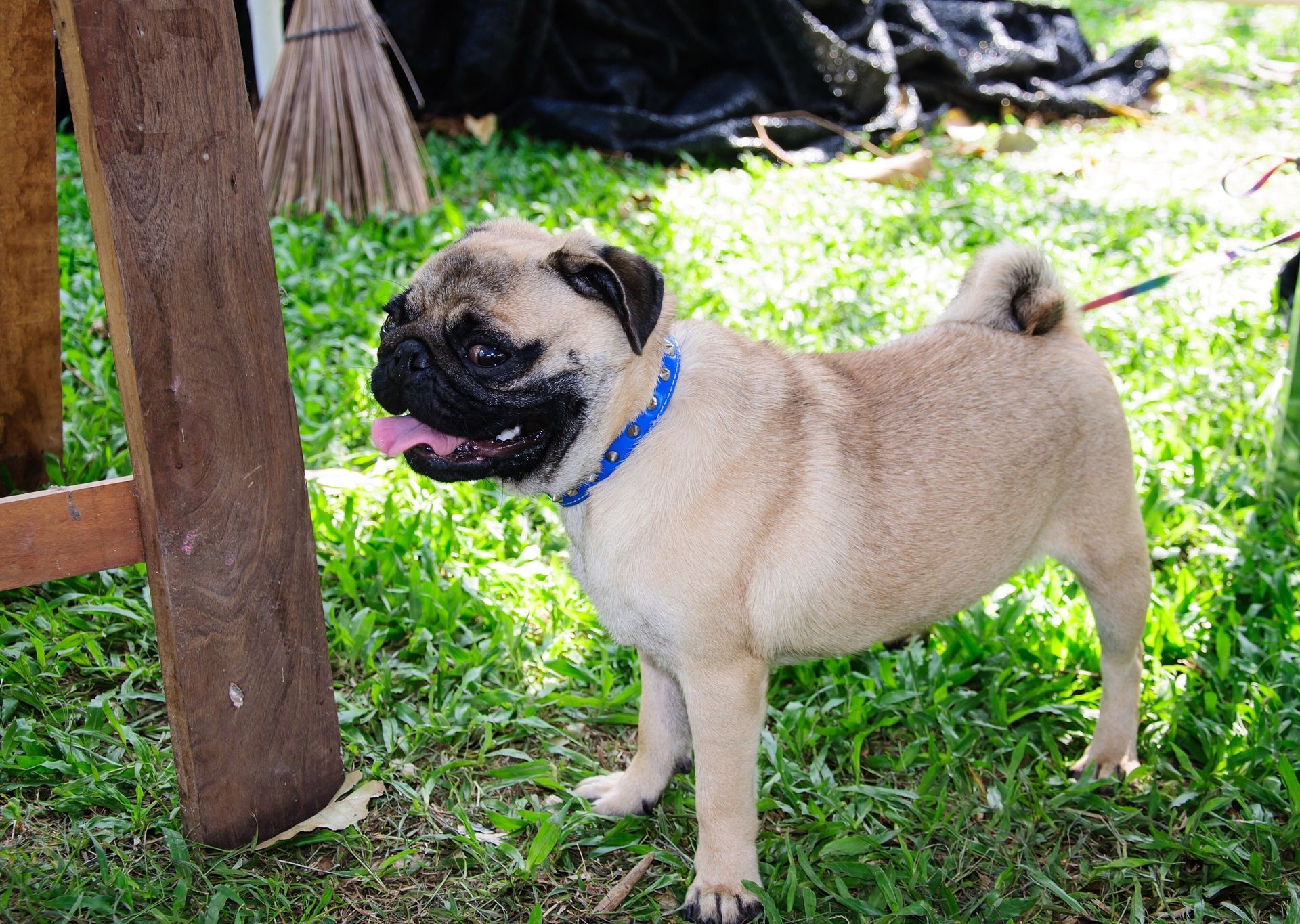
Some dog breeds face a higher risk of proptosis than others. The condition occurs most commonly in brachycephalic (flat-faced) breeds, including Pugs, Boston Terriers, and French Bulldogs. The foreshortened faces and large eye sockets of these animals make eyeball protrusion more likely.
What Should You Do If Your Dog Has Proptosis?
If your dog shows signs of proptosis, do not try to push the eye back into its socket. Instead, contact a veterinary center immediately for professional advice and to let them know of your imminent arrival. Bring your dog to the veterinary clinic as soon as safely possible.
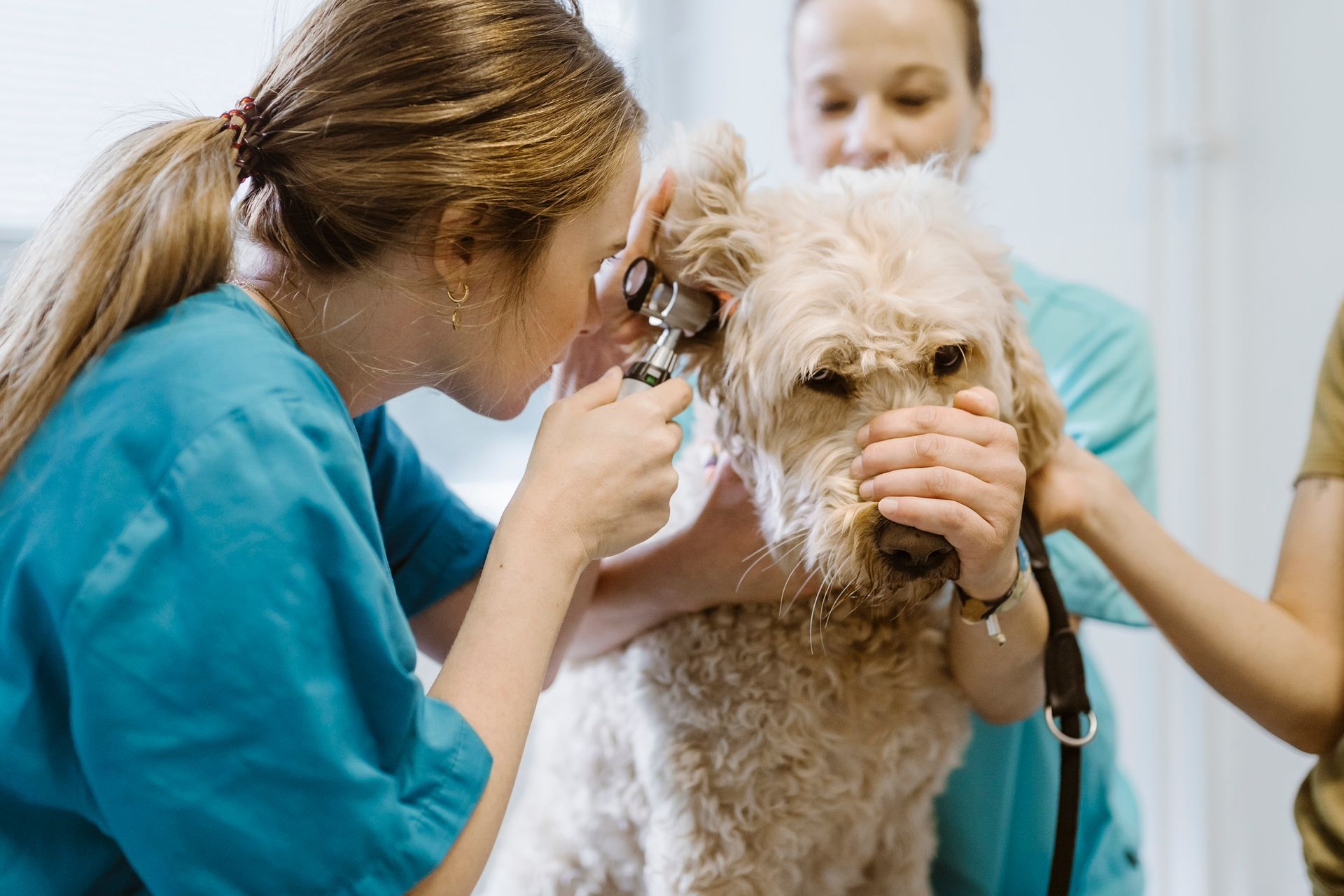
The veterinary team will evaluate the state of the eyeball and eye socket carefully. If the affected eye still responds to bright light, the eye still functions and may preserve at least some vision. The attending veterinarian will also examine the outer eye structures for ulceration, bleeding, and other trouble signs.
How Do Veterinarians Treat Canine Proptosis?
If the retina, optic nerve, and most of the eye muscles remain intact, the veterinarian may administer general anesthesia, clean and lubricate the tissues, and then try to restore the eyeball to its socket. Sutures then hold the eyeball in place during the healing process. Recuperation includes a schedule of medication and evaluations.
Over 60 percent of proptosis-afflicted eyeballs suffer too much damage to preserve vision or make restoration practical. If your dog faces this scenario, your veterinarian will surgically remove the eye, a procedure called enucleation. Fortunately, dogs who undergo enucleation adjust to this loss relatively easily.
Whether your dog needs immediate eye treatment or routine wellness evaluations, Baywood Animal Hospital can put your mind at ease by providing a full range of veterinary services. Contact our office for emergency advice or to schedule an appointment.

